
Figure 1.
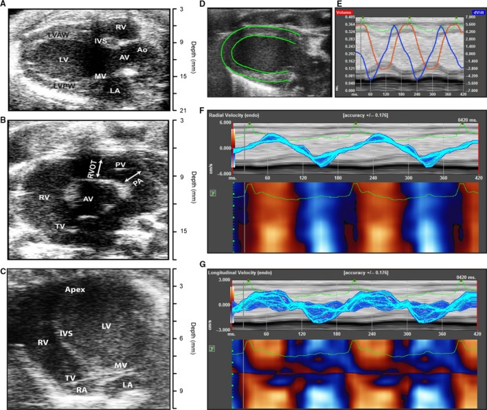
Echocardiographic analysis of the heart. (A) Parasternal long‐axis view of the heart. (B) Short‐axis view of the heart. (C) Apical four‐chamber view of the heart. (D) Tracing the left ventricular myocardial borders for strain analysis. (E) Strain analysis indicating left ventricular volume (red) and derivative of tissue velocity (blue) wall motion in two cardiac cycles. The green line indicates the corresponding EKG. (F) Radial strain for two long‐axis LV contractions. (G) Longitudinal strain for two long‐axis LV contractions. Ao, aorta; AV, aortic valve; IVS, interventricular septum; LA, left atrium; LV, left ventricle; LVAW, left ventricular anterior wall; LVPW, left ventricular posterior wall; MV, mitral valve; PA, pulmonary artery; PV, pulmonic valve; RA, right atrium; RV, right ventricle; RVOT, right ventricular outflow tract; TV, tricuspid valve.