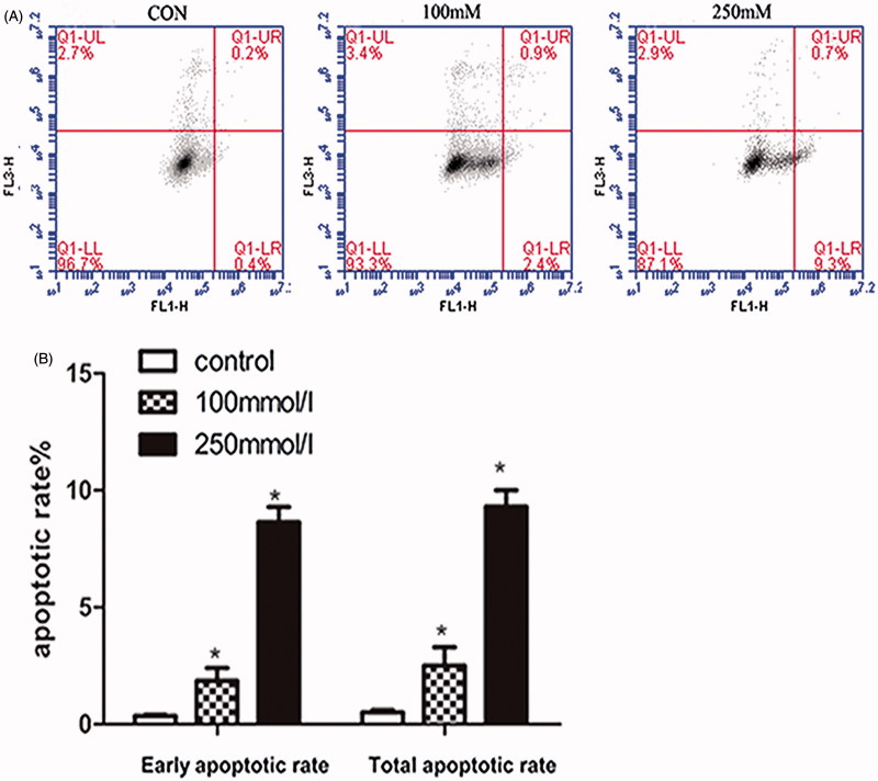
Figure 3.
Effects of mannitol on the apoptosis of HK-2 cells. Flow cytometry assays showed apoptosis of HK-2 cells treated with 0, 100, 250 mM mannitol for 48 h. (A) Shown are representative dot plot of cells stained with Annexin V-FITC/PI following treatment with mannitol for 48 h. (B) Bars represent the cell percentages of early and total apoptotic cells after treatment with different concentrations of mannitol. Note: Compared with the control group, ‘*’ indicated significant difference (p < .05).

