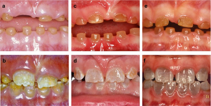
Figure 3.

Clinical photographs of the primary and permanent teeth of the probands. Left row: Intraoral photographs of Case A‐II.2 (A: 6 years; B: 7½ years). Middle row: Intra oral photographs of Case B‐III.2 (C: 3½ years; D: 7½ years). Right row: Intraoral photographs of Case C‐III.2 (E: 3½ years; F: 9 years). Note the similar amber brown color of the primary teeth in the all cases (a–c), whereas there is a marked difference in color of the permanent teeth; in Case A the discolored teeth are yellow brown (b) Case B the discoloration is light‐brown (d), and in Case C it is grayish‐blue (f)
