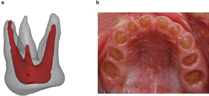
Figure 4.

(a) 3D picture of an extracted maxillary first molar in the primary dentition of Case B‐III.2 at the age of 2 years. The size of the enlarged pulp chamber is illustrated. (b) Intraoral photograph of Case C‐IV.1 at the age of 2.5 years exemplifying the excessive attrition with pulp exposure in DI–II patients
