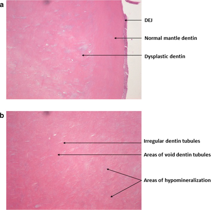
Figure 5.

The histological analysis of the teeth in Case B‐III.1 revealed the presence of (a) A dentino‐enamel junction with an even appearance suggesting lack of scalloping, a normal mantle dentin, and an underlying layer of dysplastic dentin with incomplete mineralization, recognized as pale or blue staining areas. (b) The dentin tubules were irregular and enlarged with frequent branching in the circumpulpal dentin in the crown
