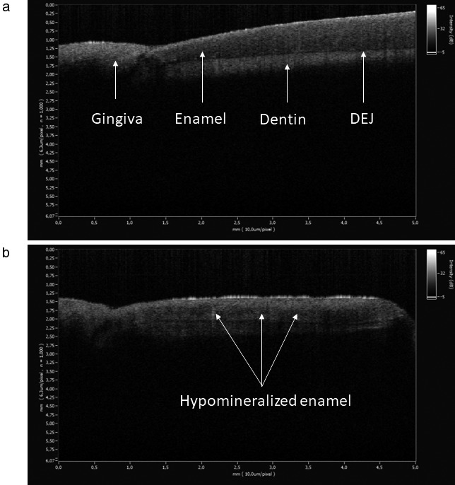
Figure 6.

Imaging results of 2‐ from the author K.T and from Case B‐III.2. (a) The healthy enamel had a normal homogeneous appearance. (b) The dysplastic enamel was characterized by dark horizontally arranged areas of hypomineralization adjacent to the dentino‐enamel junction
