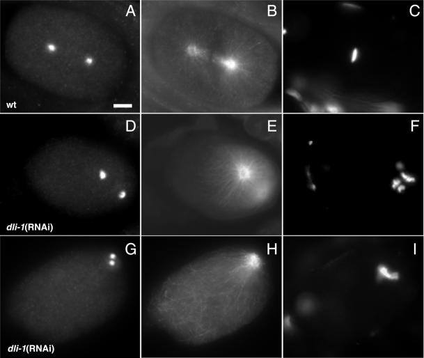
Figure 6.
DLI-1 is required for centrosome separation in the one-cell C. elegans embryo. Cytological analysis of metaphase embryos stained for the centrosomal component ZYG-9, tubulin, or DNA as indicated. All images are oriented with anterior to the left. (A–C) Wild-type embryo at metaphase. (A) Anti-ZYG-9 stains the two centrosomes that have rotated and aligned along an anterior-posterior axis. (B) Centrosomes nucleate microtubules forming the bipolar spindle visualized by anti-tubulin staining. (C) DAPI staining shows chromosomes condensed to the metaphase plate. (D–F) dli-1(RNAi) embryo in which centrosomes did separate. (D) Centrosomes have separated but have failed to centrate. This panel represents the most successful degree of rotation and spindle alignment observed. (E and F) Centrosomes do nucleate microtubules and chromosomes are condensed but have not yet congressed to a metaphase plate. (G–I) dli-1(RNAi) embryo exhibiting the most severe centrosome separation phenotype. (G) Some embryos (10/17) from wild-type injections failed to separate centrosomes and therefore formed no bipolar spindle. (H) Microtubules are nucleated and chromosomes have condensed and congressed to a metaphase plate (I). Embryos from heterozygous injections (7/8) were phenotypically identical to the embryo in G–I. Bar, ∼10 μm.

