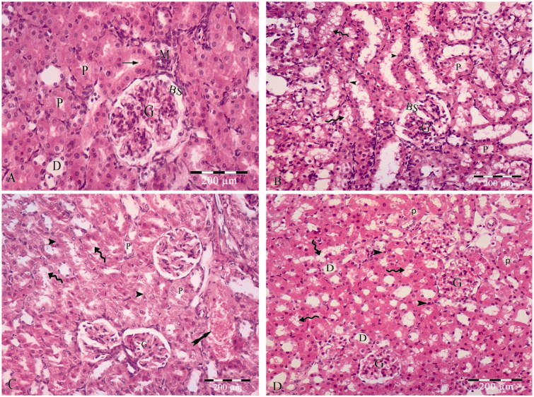
Figure 1.
(A) photomicrograph of hematoxylin and eosin stained paraffin sections; (A) Control rat renal cortex showing glomerular capillary (G), Bowmans space (BS), proximal convoluted tubules (P) lined with cuboidal cells with acidophilic cytoplasm, rounded open face nuclei and apical brush border (arrow), distal convoluted tubules (D) and macula densa (M). (B) The sections of group II show apparent widded Bowmans space (BS) of the glomerulous (G). The cell linning of the proximal convoluted tubules (P) with lost apical brush border, apoptotic linning (arrow heads) cells having small darkly satined nuceil and slighly eosophillic vaculated cytoplasm (curved arrows). (C) The sections of group III shows of the glomerulous (G). The cell linning of the proximal convoluted tubules (P) is distorted with lost apical brush border, apoptotic linning (arrow heads) cells having small darkly satined nuceil and slighly eosophillic vaculated cytoplasm (curved arrows). Areas of haemrrage are also detected (tailed arrow). (D) Section in zinc treated renal cortex demonstrates apparently normal glomerular capillaries (G) and minimal vaculation in proximal tubular (P) lining cells (curved arrows) with small darkly stained nuclei (arrow heads). The distal convoluted tubules (D) appears normal.

