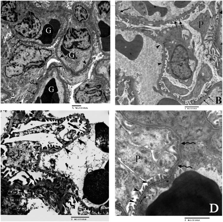
Figure 2.
A) An electron micrograph demonstrating the renal glomerular corpuscle of the control rat with a part of the cell body of podocytes (P) and glomerular capillaries (G). The filtration barrier is formed of minor processes of the podocytes (arrows), fenestrated endothelium of the glomerular capillaries (arrow heads) separated by uniformly thick basement membrane (curved arrows). B) An electron micrograph of the renal corpuscle of group II rats showing irregular minor podocytic processes (arrows) and areas of markedly thickened basement membrane (curved arrows). Arrow heads point to the irregular fenestrated endothelium. C) An electron micrograph of the renal corpuscle of group III rats showing a part of the body of podocte (P) with irregular minor podocytic processes (arrows) and areas of markedly thickened basement membrane (curved arrows) and irregular fenestrated endothelium (arrow heads). D) An electron micrograph of the renal corpuscle of group IV rats demenestrating slight thickneneing of the basement memebrane (curved arrows) and regular minor processes (arrow heads) of podocytes (P).

