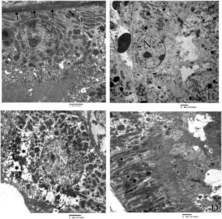
Figure 3.
A) An electron micrograph of the lining cells of proximal tubules of control rat with thick basement membrane (arrows), rounded euchromatic nucleus (N) with prominent nucleolus (n), numerous mitochondria (M) with regular basal infolding (tailed arrow) and apical microvilli (V). B) An electron micrograph of proximal tubules of group II rats showing, area of microvilli (V) with some areas of detachment, swollen bizarre shape mitochondria (M), secondary lysosomes (L) and normal nucleus (N). C) An electron micrograph of proximal tubules of group III rats showing, area of detached microvilli (curved arrow), swollen bizarre shape mitochondria (M), wide cytoplasmic spaces (stars) multiple secondary lysosomes (L) and apparently normal nucleus (N). D) An electron micrograph of the lining cells of proximal tubulus of group IV rats showing regular elongated mitochonderia (M), secondary lysosomes (L) with normal basal infolding (tailed arrow) and normal apical microvilli (V).

