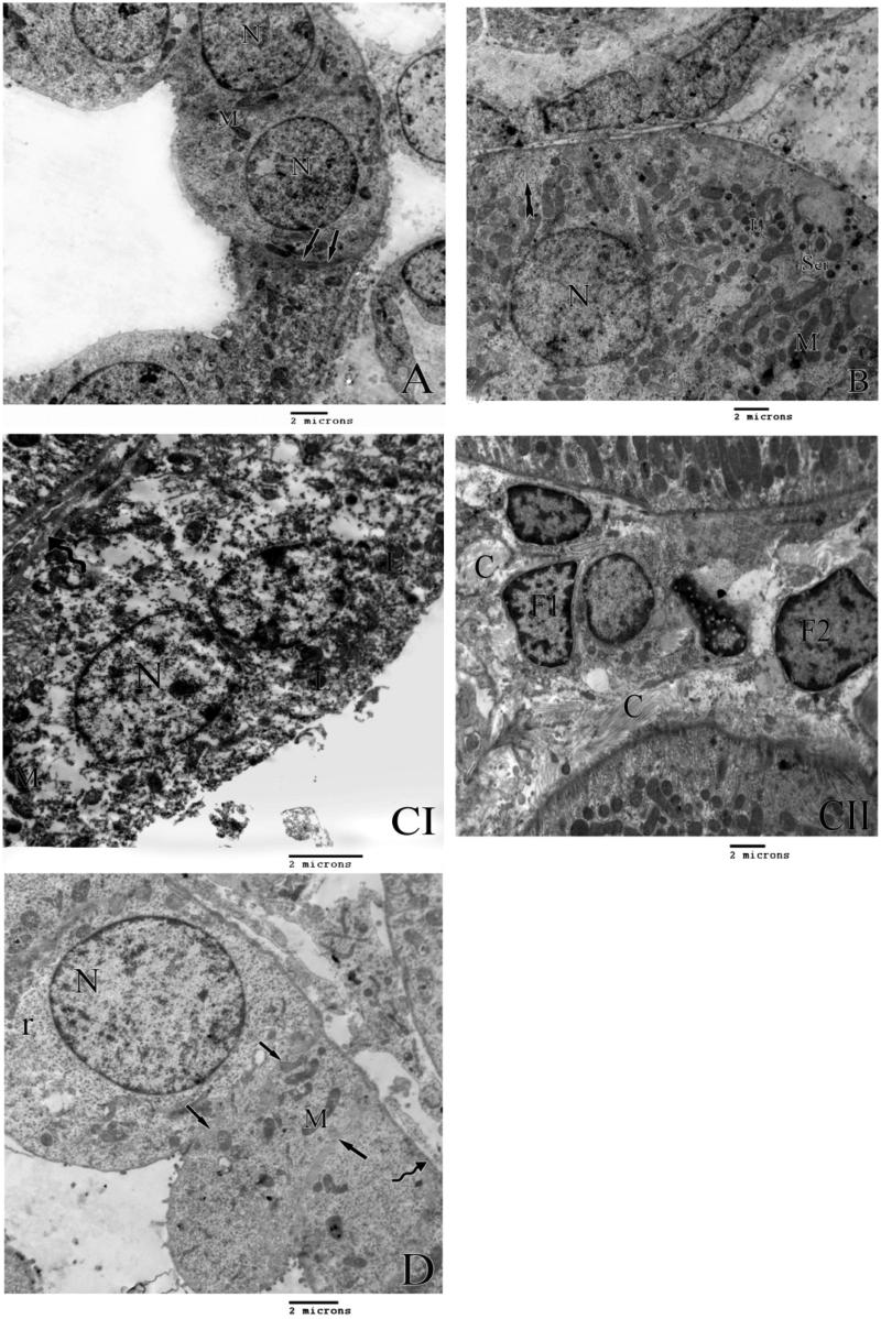
Figure 4.
A) An electron micrograph of the control rat distal tubule lining cells showing definite cell border (arrows) and elongated mitochondria (M) and rounded nuclei (N). B) An electron micrograph of the lining cells of the distal tubules of group II rats showing distorted basal enfolding (tailed arrow) bizarre shape mitochondria (M) swollen smooth endoplasmic reticulum (Ser) secondary lysosomes (L) and rounded nucleus (N). C) An electron micrograph of the lining cells of the distal tubules of group III rats showing irregular thickening of the basement membrane (curved arrow), bizarre shape mitochondria (M), secondary lysosomes (L) and rounded nucleus (N). II) shows areas of fibrosis in the interstitial tissue (C) with primary and secondary fibrocytes (F1 and F2 respectively). D) An electron micrograph of lining cells of distal tubules of group C rats showing rounded nucleus (N) numerous free ribosomes (r) with distict cell border (arrows), normal mitochonderia (M) with normal thickened basement membrane (curved arrow).

