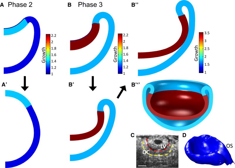
Fig. 10.
Simulation of ECM degradation (stiffness gradient model). To simulate matrix degradation, growth is paused at a specified invagination depth (A, B) and the ECM is converted to iOV tissue by decreasing its shear modulus and increasing its growth to match those of the iOV (A’, B’). (A, A’) Phase 2 (G = 1.4 in iOV); ECM degradation causes the OV to pop out, switching from concave to convex. (B, B’) Phase 3 (G = 2.25 in iOV); the OV remains invaginated. (B”) To simulate further invagination after the lens and optic cup have detached, growth is continued after ECM degradation of B’. (B”’) 3D representation of the model in B”. (C) Phase 4 (HH17) chick optic cup (OCT cross-section) shows the model qualitatively captures the flattening at the center of the iOV. (D) Reconstruction of optic cup from OCT images shows 3D shape with a groove along the optic stalk (OS). Scale bar 100 µm

