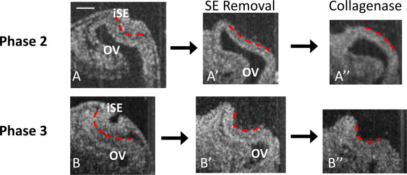
Fig. 4.
OCT images of OVs after SE removal and ECM degradation. Dashed red lines indicate outer surface of iOV. (A, A’, A”, HH14−) After removing the SE during early invagination, the OV sometimes remained concave (A’, n = 10/34 concave), but exposure to collagenase usually caused the remaining concavity to reverse (A”, n = 5/6 convex). (B, B’, B”, HH14+) At later phases of invagination, the OV typically remained concave after lens removal (B’, n = 25/40 concave) and usually remained concave after subsequent collagenase exposure (B”, n = 5/14 convex). At stages with a more developed optic cup (~HH15, not shown), concavity was maintained in all embryos after SE removal (n = 15/15 concave) and ECM degradation (n = 4/4 concave). Scale bar 100 µm

