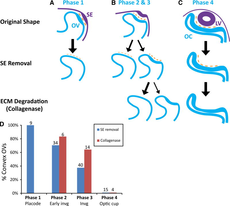
Fig. 5.
Summary of shape changes in OV after SE removal and ECM degradation. A–C Schematics of observed shape changes (purple = SE, blue = OV, orange = ECM). Arrow size reflects the likelihood of each shape change as plotted in (D). (A, Phase 1) Removing the SE at the placode stage prior to invagination caused relatively little immediate shape change. (B, Phases 2 and 3) During OV invagination (HH14− to HH14+), SE (lens placode) removal sometimes caused the OV to lose its concave shape and become convex. Exposing the remaining concave OVs to collagenase caused most to pop out and become convex. (C, Phase 4) At later stages of invagination (about HH15 onward), lens removal and subsequent collagenase treatment had relatively little effect on the shape of the OC. (D) Fraction of OVs that became convex after treatment. As invagination deepened, optic cups were more likely to remain concave after SE removal (P < 0.001, Fisher’s exact test comparing Phases 2 and 3). However, collagenase had a similar effect at these two phases (P < 0.613, Fisher’s exact test). Numbers above bars indicate sample size. All OVs remained concave at Phase 4 after both SE removal and collagenase treatment

