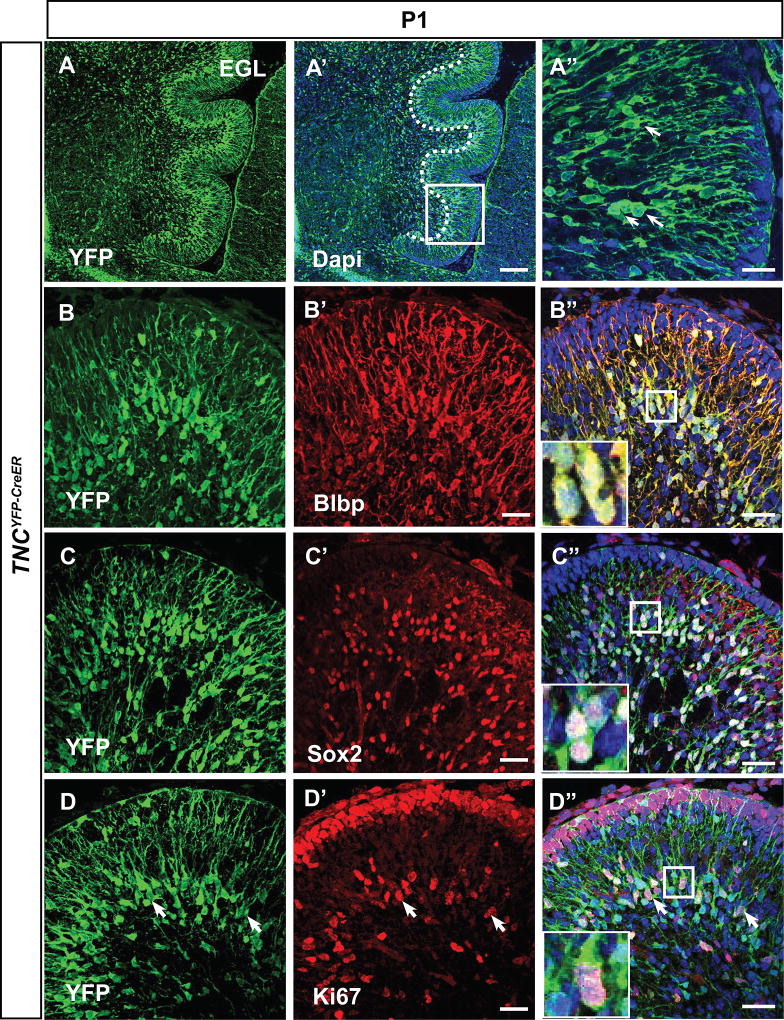
Figure 1. TNC-YFP-expressing cells are Bergmann glia.
(A–A”) YFP immunohistochemistry on sagittal sections demonstrates pattern of TNC expressing cells. Boxed region denotes enlarged area in (B’). TNC-YFP expression is observed in cells that extend long processes to the pial surface (A”). (B–C”) TNC-YFP expressing cells are Blbp+ (B–B”) and Sox2+ (C–C”), indicating that they express astroglial markers. Inset shows example of co-labeled cell (B”, C”). (D–D”) TNC-YFP expressing cells are ki67+, indicating that they are proliferative. Inset shows example of co-labeled cell (D”). Abbreviation: EGL, external granular layer. Scale bars are 20 µm except A”, B”, C” and D” (100 µm).

