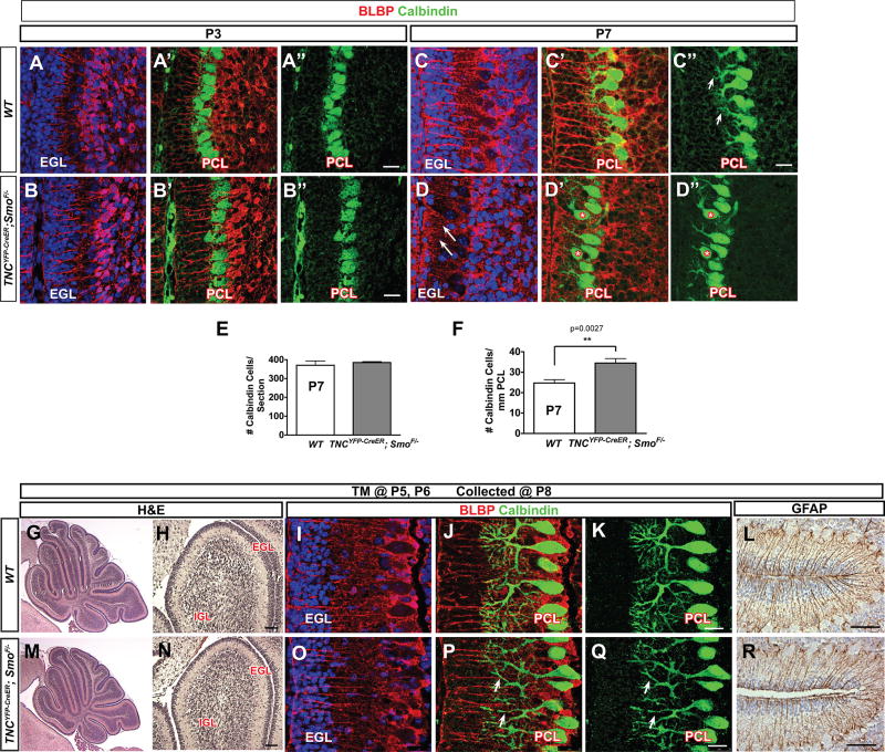
Figure 7. Smocko mutants have disrupted alignment and dendritic arborization of PCs.
(A–B”) At P3, Calbindin immunohistochemistry shows no difference in Smocko mutant (B–B”) PC morphology, layering, or dendritogenesis. (C–C”) At P7, Calbindin immunohistochemistry shows that PC soma localization is disrupted and have a severely disrupted fiber network, with stunted, thinned dendrites and poorly branched arbors. (E) The absolute number of Calbindin+ PCs were comparable between the WT and mutant. (F) There was a significant increase in the number of PCs per mm of PCL in Smocko mutants. Data are mean of n=3 WT and littermate pairs for each genotype. (G–R) Tamoxifen injection scheme in Smocko mutant at later timepoints. One dose of tamoxifen was injected at P5 and P6 in WT (G–L) and Smocko mutants (M–R). Mice were analyzed at P8. (G, H, M, N) H&E staining shows that changes in cerebellar size and EGL area were less severe when BG Shh signaling was ablated at P5 and P6 compared to ablation at P1 and P2. (I–L, O–R) PC dendrites in later timepoint-injected Smocko mutants (O–Q) lacked secondary branching structures as revealed by Calbindin immunohistochemistry (arrowheads) and no appreciable differences in BG fibers can be observed using Blbp (I–K, O–Q) and GFAP (L, R) immunohistochemistry. Abbreviations: EGL, external granular layer. IGL, internal granular layer. PCL. Purkinje cell layer. Scale bar: 100µm and 20 µm.

