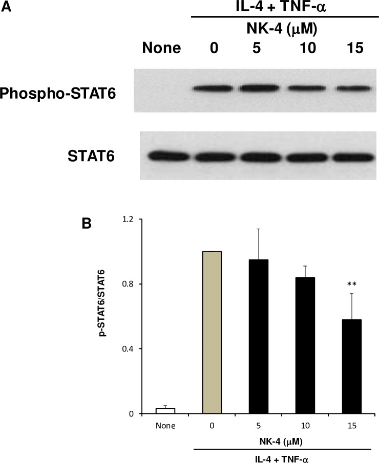
Fig 9. NK-4 suppresses the STAT6 signaling pathway in NHDF stimulated with IL-4 and TNF-α.
NHDF were grown in confluent monolayer cultures in 12-well plates and were stimulated with 10 ng/ml IL-4 and 5 ng/ml TNF-α in the presence or absence of varying concentrations of NK-4 for 15 min. Phosphorylation of STAT6 was determined by immunoblotting whole-cell lysates using specific antibodies against the phosphorylated or total STAT6 protein. A representative blot is shown (A). The optical density ratio of phospho-STAT6 to total STAT6 is shown (B). Data from three independent experiments were combined and expressed as the means ± SD. **p < 0.01 compared with control cultures. Original uncropped and unadjusted Western blots were provided as supplementary files (S1 Fig and S2 Fig).

