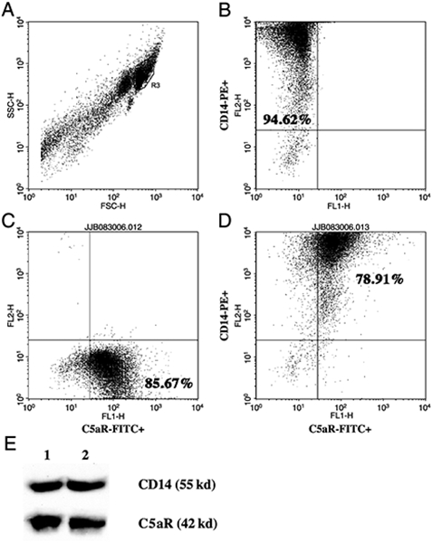
Fig. 1. The C5a receptor (CD88) is present at the surface of human PBMCs.

A–D, Flow cytometric analysis of adherent PBMCs treated with C5a for 20 h from a representative healthy volunteer. A, Forward scatter/side scatter of peripheral blood leucocytes. Cells in the R3 region were regarded as CD14+ monocytes. B, Representative dot blot depicting log fluorescence of FL-2 (CD14-PE) on the y axis and FL-1 (FITC) on the x axis. Cells in the upper left quadrant are CD14-PE+. The percentage of positive cells represents the mean of 3 independent experiments. C, Representative dot blot depicting log fluorescence of FL-2 (PE) on the y axis and FL-1 [C5aR (CD88)-FITC] on the x axis. Cells in the lower right quadrant are C5aR (CD88)-FITC+. The percentage of positive cells represents the mean of 3 independent experiments. D, Representative dot blot depicting log fluorescence of FL-2 (CD14-PE) on the y axis and FL-1 [C5aR (CD88)-FITC] on the x axis. Cells in the upper right quadrant are CD14-PE+ and C5aR (CD88)-FITC+. The percentage of positive cells represents the mean of 3 independent experiments. E, Western blot of adherent PBMCs untreated (lane 1) or treated with C5a (100 ng/mL) for 20 h (lane 2). Membranes were probed with anti-CD14 and anti-C5aR (Cd88) antibodies.
