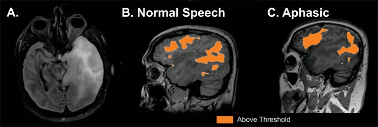
Fig 5. Resting state MRI mapping of speech networks in an aphasic patient.
A. Forty year old patient with left temporal tumor. B. rs-fMRI mapping of speech when speech was intact. C. rs-fMRI mapping of speech when patient globally aphasic due to prolonged seizure. Imaging threshold for MLP rsfMRI was 97% probability.

