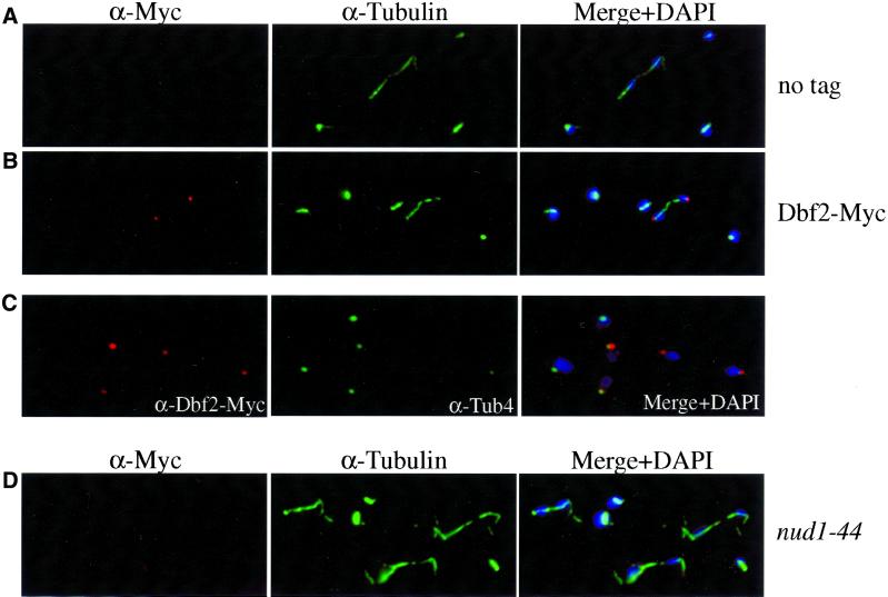
Figure 2.
The subcellular localization of Dbf2-Myc during the cell cycle. (A) Exponential growing cells lacking a Dbf2-Myc fusion (no tag; K699). (B) Dbf2-Myc and microtubules of strain A1931 were visualized with the use of anti-Myc antibodies (BABCO) and antitubulin antibodies (α-tubulin), respectively. DAPI was used to stain DNA (DAPI). (C) Colocalization of Dbf2-Myc with the spindle pole body component Tub4. (D) Dbf2-Myc localization in nud1–44 mutant (A2340) 120 min after shift to the restrictive temperature (37°C).

