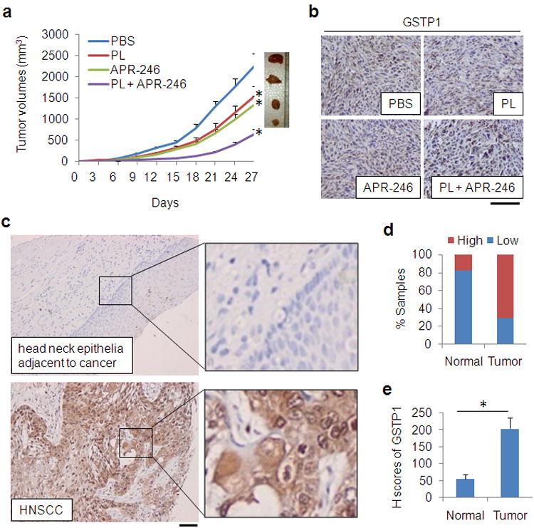
Figure 7. GSTP1 is highly expressed in HNSCC tissues. Application of PL and APR-246 reduces xenograft tumor growth of HNSCC cells.

(a, b) 5 × 10⁶ UMSCC10A cells were inoculated subcutaneously under the flank of SCID mice. Three days later, the mice were randomized into4 groups (n = 10 / group) receiving PBS (control), PL (4 mg/kg/day), APR-246 (100 mg/kg/day), or PL (4 mg/kg/day) + APR-246 (100 mg/kg/day) by i. p. Treatment was performed daily for 24 days. (a) Tumor volumes were measured every 3 days. *P < 0.01 as compared with control treatment group. (b) The tumors were removed from euthanized mice. IHC was used to detect GSTP1. Scale bar = 100 μm. (c - e) HNSCC tissues from healthy (n = 28) and HNSCC (n = 194) subjects were assessed for the expression of GSTP1 by IHC. (c) Representative IHC staining of GSTP1 in a normal head and neck epithelial tissue and in an HNSCC tissue. Scale bar = 100 μm. (d) Quantification of GSTP1 expression in human head and neck tissues. Low: overall negative or weak staining; High: overall moderate or strong staining. The Pearson's chi-square test was used to analyze the distribution difference of GSTP1 between healthy and HNSCC tissues (P < 0.01). (e) H-scores of GSTP1 in head and neck tissues (*P< 0.01).
