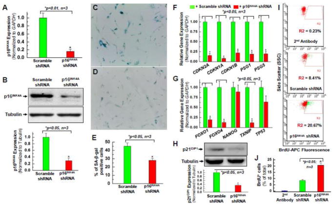
Figure 2. Knocking down the expression of p16INK4A reverses the senescent phenotype of hCPCs.
Real time qPCR data (A) and quantitative analysis of Western blot (B) confirmed that p16INK4A protein expression was knocked down in one line (AMC20) of the hCPCs-A group at passage 8 (P8) infected with lentivirus expressing shRNA against p16INK4A compared to the control which was infected with scramble shRNA. Senescence-associated β-galactosidase staining showed a decrease in the number of senescent cells in the group of hCPCs that were infected with lentivirus expressing shRNA against p16INK4A (D) compared to the group of hCPCs expressing the scramble shRNA (C). E. Quantitative analysis of β-galactosidase positive staining in panel C and D. In hCPCs with 48-hour knockdown of p16INK4A, senescence-associated genes (F) and quiescence-associated genes (G) exhibited decreased expression compared to hCPCs with 48-hour infection by a scramble control. Data labels shown are for the gene expression levels in p16INK4A shRNA-infected hCPCs, relative to those in scramble shRNA-infected hCPCs. H. Quantitative analysis of Western blot confirmed a decrease in p21CIP1 protein expression after knocking down p16INK4A in hCPCs, consistent with its decreased gene expression (CDKN1A) shown in panel F. I. Representative FACS analysis showed the increased proliferation ability of hCPCs after knocking down p16INK4A. J. Quantitative data analysis for panel I. Data were presented as means ± SD from 3 independent experiments (n=3); * indicates p<0.05 vs. scramble shRNA.

