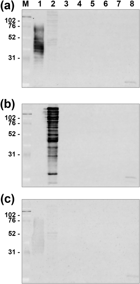
Figure 1.

Western blot analysis for reactivity of the anti-PG and anti-TF antibodies. Western blot analysis for reactivity of the anti-PG antibody (a; upper column), the anti-TF antibody (b; middle column), and the PBS control without primary antibody (c; lower column) was performed using sonicated whole-cell lysate. M; marker of molecular weight (molecular mass in kiloDaltons), Lane 1; PG (ATCC 33277), lane 2; TF (ATCC 43037), lane 3; Aggregatibacter actinomycetemcomitans (ATCC 43718); lane 4, Fusobacterium nucleatum (ATCC 25586), lane 5; Prevotella intermedia (ATCC 25611), lane 6; Prevotella nigrescens (ATCC 33563), lane 7; Bacteroides fragilis (ATCC 25285), and lane 8; Bacteroides vulgatus (ATCC 25285). Both the anti-PG and anti-TF antibodies exhibited a ladder pattern of positive bands ranging from 31 to 76 kDa and from 38 to 225 kDa only on the PG and TF lanes (a,b), respectively. No positive bands were observed in the other lanes or in the PBS control (c).
