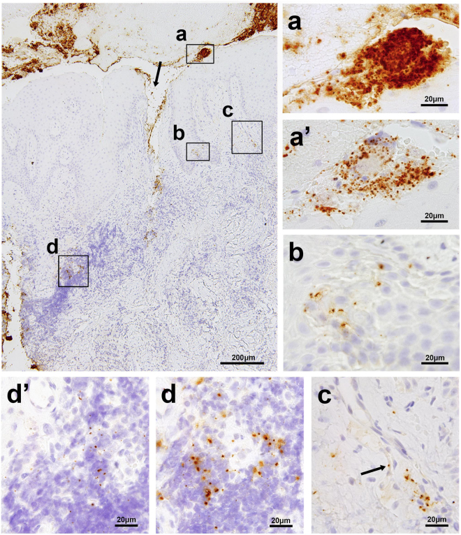
Figure 2.
Localization of PG and TF in a representative sample of gingival and subgingival tissue affected by chronic periodontitis. The largest photo shows a low-power view (original magnification, ×100) of the sample immunostained with the anti-TF antibody, representing superficially-located bacterial aggregates or plaques (a), bacterial tissue invasion through the disrupted epithelial layer (indicated by the arrow), a cluster of squamous cells with bacteria in the epidermal layer (b), a bacterium in a capillary wall of granulation tissue (c), and scattered bacterial cells in the area of heavy inflammatory cell infiltration (d). The small photos show high-power views (original magnification, ×1000) of the boxed areas indicated by (a,b,c and d), including of the photos (a’ and d’) taken from the adjacent sections immunostained with the anti-PG antibody, each corresponding to the area indicated by a and d, respectively. The arrow in photo (c) shows a single TF cell located in a capillary wall.

