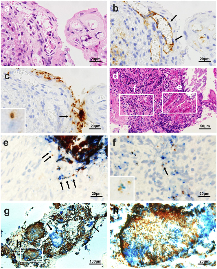
Figure 3.
Identification of a bacterium in the capillary endothelium with anti-CD31 antibody and simultaneous identification of PG and TF in identical sections. Three serial sections of a sample with chronic periodontitis with superficial capillary proliferation were stained by hematoxylin and eosin (a) and immunostained with the anti-CD31 antibody (b) and anti-TF antibody (c). The arrows indicate capillary endothelial cells (b) containing a single TF bacterial cell in the capillary endothelium (indicated by an arrow and magnified in the inset) (c). Two serial sections of a subgingival tissue sample with erosive epidermal layer and subepidermal inflammation were stained by hematoxylin and eosin (d) and immunoenzyme-double-stained with anti-PG (brown) and anti-TF (blue) antibodies (e and f). In the erosive epidermal layer (e), one or a few TF and PG cells colocalized in the epithelium (arrows). In the subepidermal inflammatory area (f), a few PG cells were found with many TF cells in some of inflammatory cells, and both species cells colocalized in an identical inflammatory cell (indicated by an arrow and magnified in the inset). A section of a sample with many bacterial aggregates and plaques was immunoenzyme double-stained with anti-PG (brown) and anti-TF (blue) antibodies (g). Note that most of dark-brown coloured signals are double-positive signals produced by overlapping of blue-coloured TF-positive signals and brown-coloured PG-positive signals. Many plaques or aggregates were intermixed with PG and TF cells, but a few comprised only TF cells (indicated by arrows). A high-power view of a bacterial plaque (h) shows the intermixture of both species with a predominant distribution of TF cells in a central core and PG cells in the marginal areas. Original magnifications: a, b, c, e, f, and h, ×1000, d, ×400, and g, ×200.

