Fig. 4.
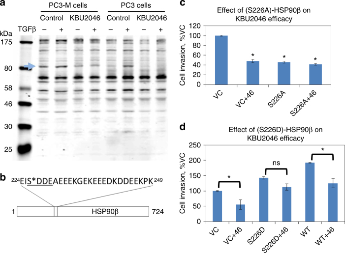
KBU2046 decreases phosphorylation of HSP90β. a Probing for KBU2046-induced changes in protein phosphorylation. PC3-M or PC3 cells were pre-treated with 10 µM KBU2046 for 3 days, then with ±TGFβ and the resultant cell lysate probed for changes in protein phosphorylation with the KinomeView® assay. The depicted western blot utilizes KinomeView® phospho-motif antibody, BL4176; the blue arrow denotes an 83 kDa band whose phosphorylation is inhibited by KBU2046 (see Supplementary Fig. 10 for complete KinomeView® assay screening data). b Proteomic analysis. PC3 cells were pre-treated with KBU2046 or vehicle, then with TGFβ, proteins from the resultant cell lysate were immunoprecipitated with BL4176, and HSP90β was identified by LC-MS/MS analysis (see Supplementary Fig. 12 for expanded proteomic assay data). The phospho-motif recognized by the antibody is underlined; S*—denotes Ser226, whose phosphorylation is decreased by KBU2046. c, d The phospho-mimetic changes in HSP90β Ser226 structure regulate human PCa cell invasion and KBU2046 efficacy. PC3-M cells were transfected with S226A-, S226D-, or WT-HSP90β, or empty vector (VC), treated with KBU2046 or vehicle, and cell invasion measured. Values are the mean ± SEM of a representative experiment of multiple experiments (all in replicates of N = 3); *denotes t-test P value <0.05 between bracketed conditions, or compared to VC

