Figure 1.
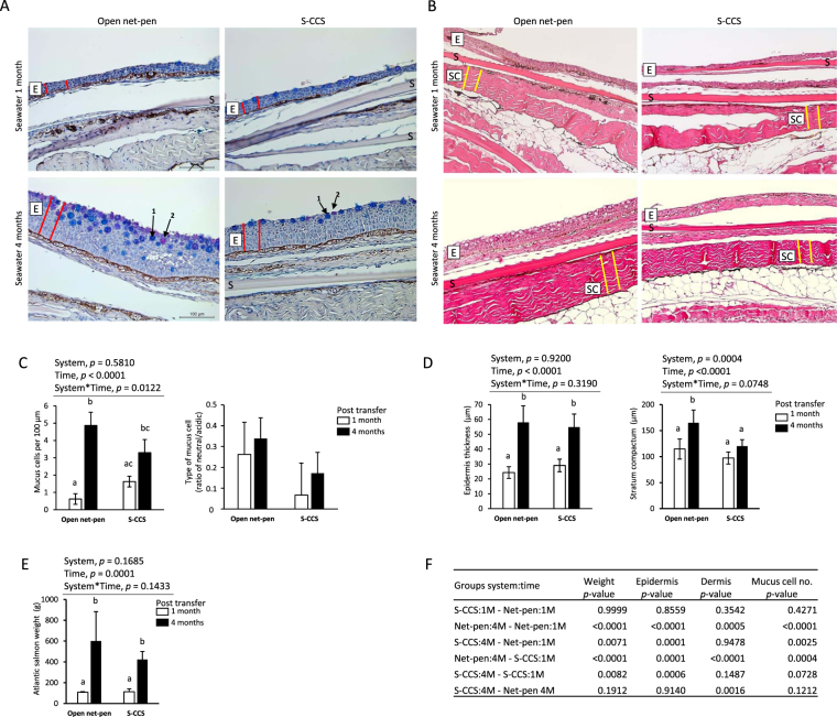
Representative images of Atlantic salmon skin 1 on 4 months post seawater transfer reared in net-pen or semi-closed containment systems (S-CCS). (A) Images displaying mucus cells assessed on alcian blue-periodic acid Stiff (AB-PAS) stained sections. AB-PAS stains the mucins in the goblet cells: acidic mucins stain blue (arrow 1) and neutral mucins stain pink-red (arrow 2). Epidermis layer indicated by a boxed E was measured by thickness (μm) as illustrated by red lines. S, scale. (B) Differences in the dermis stratum compactum (boxed SC) layer thickness (μm) was measured as illustrated by yellow lines. (C) Cells stained positive by AB-PAS were counted as goblet cells and presented as mucus cells per 100 µm. Numbers of mucus cells are different with time. The presence of different types of mucus cells is presented as the ratio between neutral and acidic stained cells. (D) Quantitative assessments (n = 10 measurements per sample) showing an increase in mean epidermal thickness between time-points but not systems. Thickness in stratum compactum of net-pen fish 4 months post seawater transfer was different to other groups. (E) Weight of Atlantic salmon used for histology sampling increased between time-points. Bars in all histograms represent the mean ± SD (n = 6 biological samples). Plots were analysed with two-way ANOVA (results in C-E above each graph where one or both variables are significant) and post hoc differences tested by Tukey HSD. Bars not sharing common letters are significantly different (p < 0.05). (F) Pairwise multiple comparisons of weight, epidermis, dermis and mucus measurements showing p-values from Tukey HSD.

