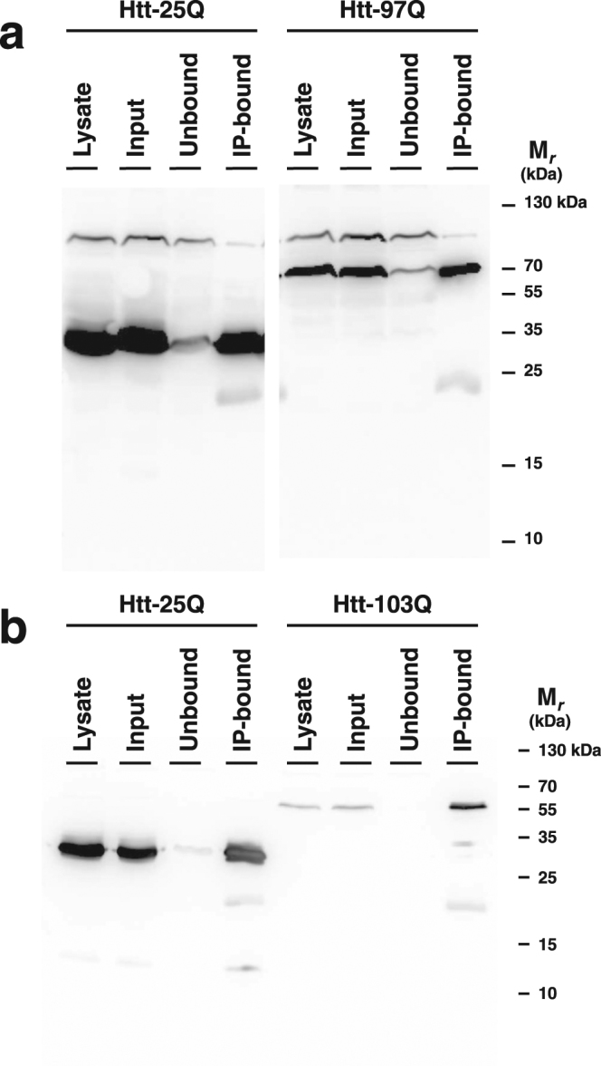
Figure 1.

Western blot of anti-FLAG immunoprecipitation. Htt was immunoprecipitated from (a) S. cerevisiae and (b) S. pombe Htt-expression strains using an anti-FLAG mAb coupled to magnetic beads, as described in Methods. Equivalent quantities (25 OD) of whole cell lysate, input, unbound, and IP-bound samples were loaded and detected with anti-FLAG mAb. Note that the two sides of panel A are cropped from different regions of the same gel. The complete gel is shown at the end of the Supplementary Information document.
