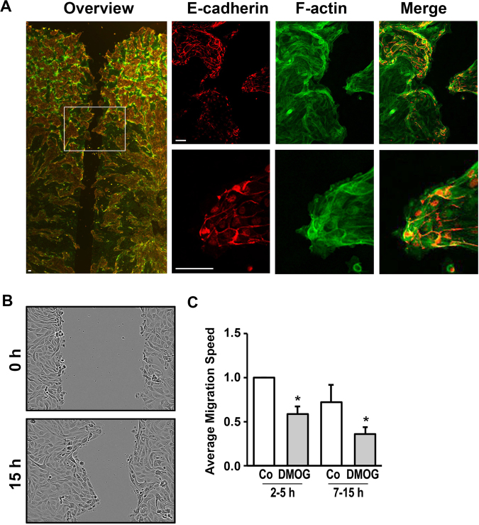
Figure 1.
Migration of hPTECs was impaired by DMOG. (A) hPTECs were seeded in Ibidi barriers in 8-well slides and grown to confluence. After removal of the barriers, cells were allowed to migrate into the open space for 7 h. Cells were stained for E-cadherin (red) and F-actin (green). Scale bar: 30 µm. (B) Distal hPTECs were seeded as described above. Pictures of the wound were taken directly after removal of the barrier and 15 h later. (C) Distal hPTECs were seeded as described above. Cells were treated with DMOG (1 mM) 24 h prior to removal of the barriers. Movement of the cells was monitored using the program provided by the manufacturer (IncuCyteR, Essen Biosciences). Based on the confluency data, migration speed was calculated in arbitrary units per h, covering an early (2–5 h) and late (7–15 h) time-frame. In each experiment, migration of control cells between 2 and 5 h was set to 1. The graph summarizes data (means ± SD) of 3 experiments. *p < 0.05 compared to control cells.

