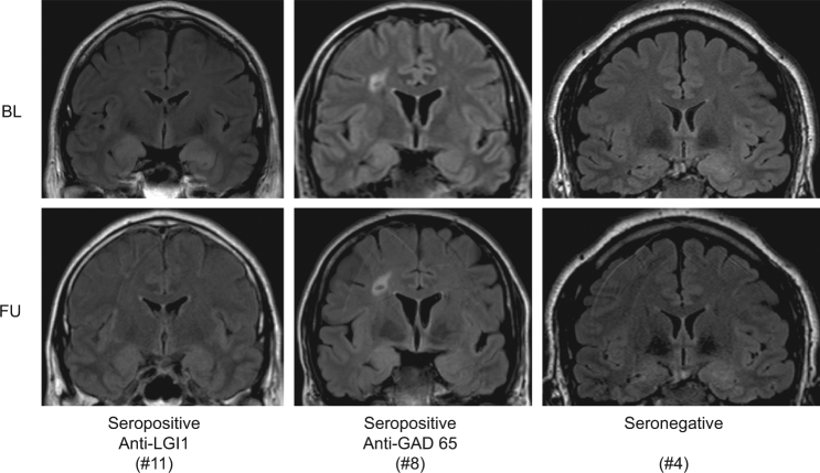
Figure 1.
Representative FLAIR images at baseline (BL, upper panels) and follow-up (FU, lower panels) of confirmed autoimmune TLE-AE with anti-LGI1 aabs (Patient #11, left panels), anti-GAD65 aabs (Patient #8, middle panels), as well as suspected autoimmune TLE-AE (Patient #4, right panels) demonstrate uni- or bilateral hyperintensity and enlargement of the amygdala. Note that changes at BL were clearly regressive at FU in patient #11.

