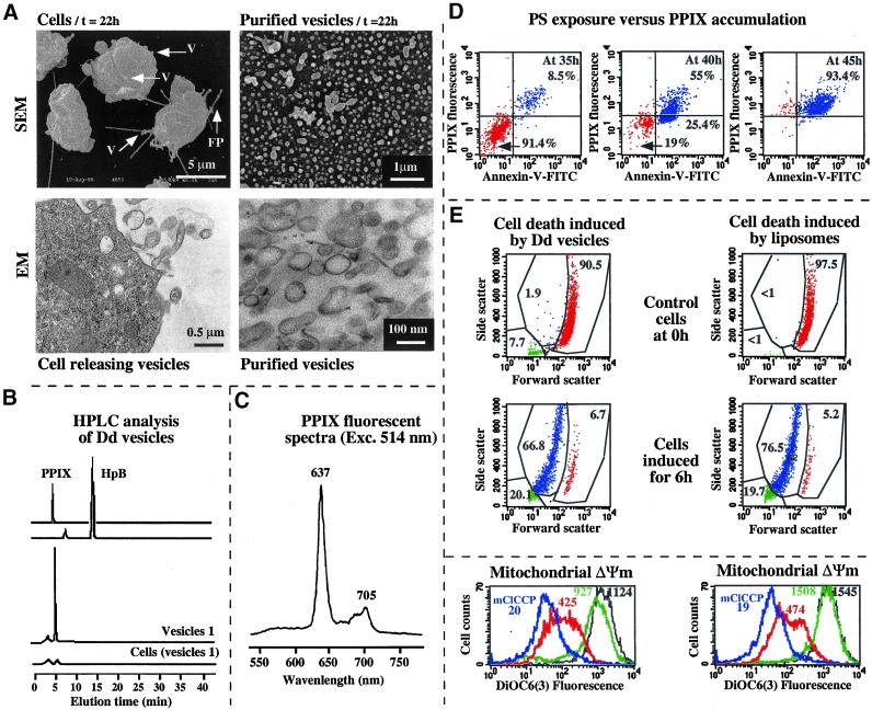
Figure 3.
PPIX: a putative inducer of Dictyostelium cell death present in the exocytotic vesicles. (a) Transmission electron microscopy and scanning electron microscopy of Dictyostelium cells releasing exocytotic vesicles and purified vesicles. V, vesicles; FP, filopodia. (b) HPLC analysis of Dictyostelium vesicles. PPIX present in cells or vesicles was quantitatively extracted (Dellinger et al., 1990). The cell or vesicle pellets were suspended in PBS (100 μl) by vigorous shaking, mixed with 2-chloroethanol (900 μl) in a glass tube and shaken gently for 30–60 min at room temperature. The extracted PPIX was stored at −20°C until dissolved in the HPLC solvent (0.2 ml) for analysis. (c) PPIX fluorescence spectra. (d) PS exposure and intracellular accumulation of PPIX in Dictyostelium. PPIX accumulation was assessed by measuring its intracellular fluorescence by flow cytometry at different times. Cell death was caused by incubation with purified exocytotic vesicles. (e) Liposomes loaded with PPIX caused Dictyostelium cell death in the same way as exocytotic vesicles with light exposure. The monoparametric histogram shows the ΔΨm after incubation with a source of PPIX and exposure to light for 15 min at 1.5 J/cm2 (in red), or with a source of PPIX without exposure to light (in black), or with source of PPIX without exposure to light (in green). Control: mClCCP, which completely abolished the ΔΨm (in blue). The results are representative of three independent experiments. HpB, hematoporphyrin B.

