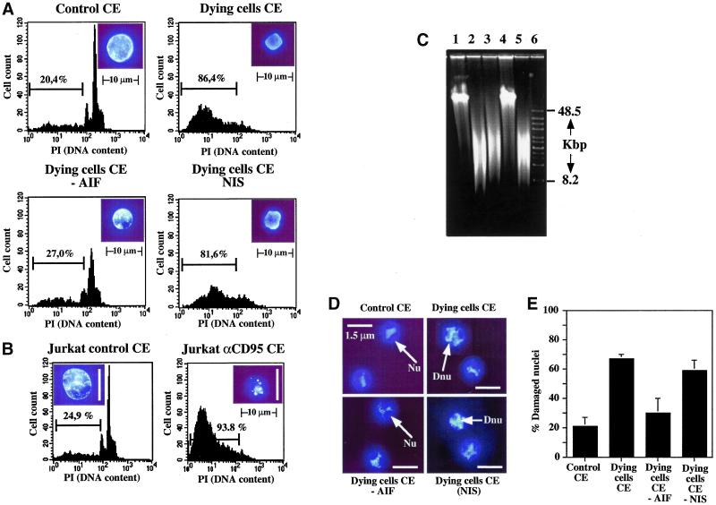
Figure 7.
DdAIF induces damage of mammalian and Dictyostelium nuclei in a cell-free system. (a) CEM nuclei were incubated for 4–5 h with cytoplasmic extracts from untreated Dictyostelium cells (control CE), or Dictyostelium cells treated with PPIX to cause cell death (dying cells CE). Cytoplasmic extracts of dying cells were also immunodepleted with anti-DdAIF (dying cells CE -AIF) or with nonimmune serum (dying cells CE NIS). Nuclei were stained with 10 μM Hoechst 33342 and examined by UV fluorescence microscopy (magnification 1000×). Nuclear apoptosis was also quantified by staining with the DNA-intercalating dye propidium iodide and analyzing the DNA content by flow cytometry. One experiment representative of three is shown. (b) Control cytoplasmic extracts were from untreated Jurkat cells or Jurkat cells incubated with 100 ng/ml anti-Fas antibody (αCD95) for 6 h at 37°C before the preparation of extracts. (c) Pulsed field gel electrophoresis of the DNA extracted from CEM cells incubated either with control CE (lane 1), dying cells CE (lane 2), dying cells CE immunodepleted with nonimmune serum (lane 3), dying cells immunodepleted with anti-DdAIF antibody (lane 4), or dying cells CE preincubated with 100 μM Z-VAD.fmk (lane 5) and HMW markers (lane 6). One experiment representative of three is shown. (d) Purified Dictyostelium nuclei were incubated for 4–5 h with the same cytoplasmic extracts as in a. The nuclei were stained with 10 μM Hoechst 33342 and examined by UV fluorescence microscopy (Leika DMRB) (magnification 2500×). (e) Quantification of damaged nuclei induced by cytoplasmic extracts. Damaged nuclei were assessed by the disappearance of the nucleoli. One hundred nuclei were counted in each condition. Histograms are the mean of three independent experiments. Nu, nucleolus; Dnu, dying cells nucleolus.

