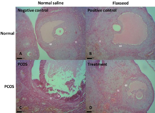
Figure 5.

A and B, healthy tertiary follicle in the negative and positive control rats. The theca layers and granulosa layers appear normal. C, A follicle in the early process of atresia with apoptotic granulosa cells, most of which are in the inner parts of the granulosa layer in a polycystic ovary syndrome (PCOS) rat. Thin and elongated epithelioid cells form the inner surface of the wall. The cyst fluid contains macrophages. D, Ovary from a flaxseed-treated PCOS rat with normal tertiary follicles. Gl, granulosa layer; Tl, theca layer (H&E staining; index bars, 50 μm)
