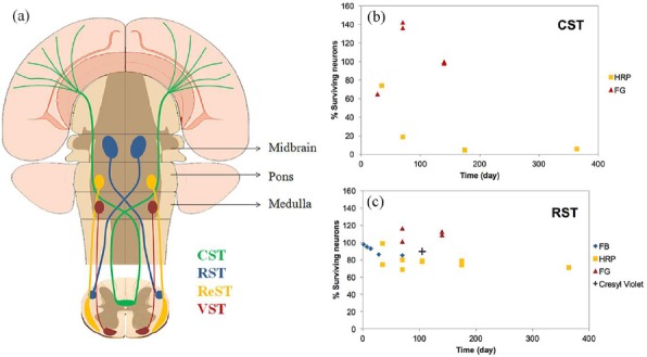
Figure 3.

(a) Schematic representation of the course of 4 most studied descending tracts including corticospinal tract (CST), rubrospinal tract (RST), reticulospinal tract (ReST), and vestibulospinal tract (VST). The percent of surviving neurons of (b) corticospinal tract (CST) and (c) rubrospinal tract (RST) following transection injury. The results are categorized based on the evaluation methods including tract tracing by FG, HRP, FB () and cresyl violet staining (+)
