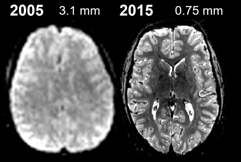
Fig. 2. Recent progress in fMRI spatial resolution and image quality.

(Left) A standard 3 Tesla 3.1 mm isotropic echo-planar image used for conventional fMRI studies circa 2005. (Right) A more recent example of 7 Tesla 0.75 mm isotropic echo-planar image, acquired with parallel imaging acceleration and a 32-channel RF coil array and a specialized head gradient coil to achieve a four-fold accelerated rapid readout. Continued technology development will be needed to surpass this resolution for whole-brain neuroimaging studies.
