
Figure 3.
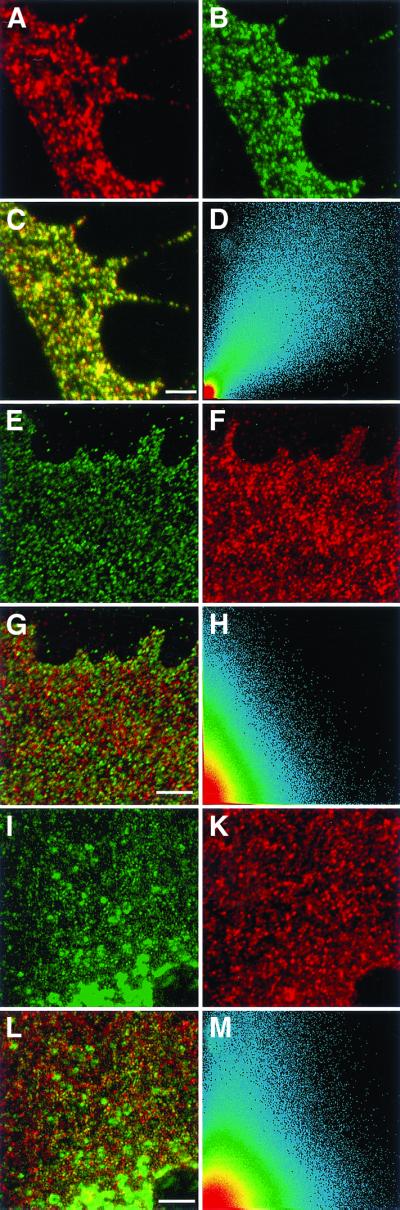
Colocalization of reggie-1 and reggie-2 and lack of colocalization with caveolin-1 in astrocytes. Optical section through processes of astrocytes showing reggie-1 (a, red) and reggie-2 (b, green) staining at the plasma membrane. (c) Superposition of the two images and the scatter plot (d) indicate substantial colocalization of reggie-1 and -2. After staining with anti-reggie-1 (e, green) and anti-caveolin-1 (f, red), or with anti-reggie-2 (i, green) and anti-caveolin-1 (k, red), the red and green dots remain separate (g and l) when the images are merged, which is also reflected by the scatter plots (h and m). Bars, 2 μm (c); 2 μm (g); 5 μm (1).