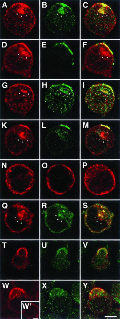
Figure 7.
Capping-induced translocation of reggie in Jurkat cells. In stimulated Jurkat cells, anti-reggie-1 (a, red) and anti-reggie-2 (b, green) accumulate preferentially at one aspect of the cell where they show substantial colocalization in patches (c, yellow). Reggie-1 (d, red) exhibits a substantial degree of colocalization with Thy-1 (e, green) as reflected by the merged images (f, yellow), and so does reggie-1 (g, red) and fyn (g, green) as seen in i, and fyn (k, red) and Thy-1 (l, green) as seen in m. Optical sections of unstimulated Jurkat cells show the unpatched distribution of reggie-1 (n), Thy-1 (o), and fyn (p), and of reggie-1 (q) and reggie-2 (r and s). The white arrowheads mark the intracellular accumulation of the respective antigens in endolysosomal compartments. Capping of Thy-1 by AB cross-linking (t) leads to increased phosphotyrosine staining (u), partially colocalized with Thy-1 immunoreactivity (v). Moreover, phosphotyrosine (w) and reggie-2 pAB immunostaining (x) partially colocalize (y) in cells stimulated by cross-linking of Thy-1. The inset (w′) shows the distribution of phosphotyrosine in unstimulated cells. Bars, 5 μm.

