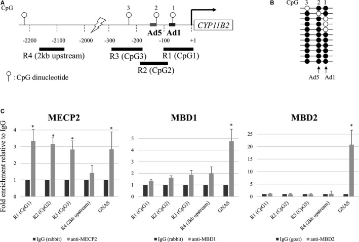
Figure 6.

In vivo interactions of methyl CpG binding domain (MBD) proteins with the CYP11B2 promoter (chromatin immunoprecipitation assay). A, Schema of the CYP11B2 promoter. B, CpG methylation status of CpG1, CpG2, and CpG3 in H295R cells. C, Recruitment of MBD proteins. R1, R2, and R3 denote regions 1, 2, 3 amplified to measure MBDs interaction with CpG1 (Ad1), CpG2 (Ad5), and CpG3, respectively. R4 was used as a control that contained no CpG site. Primers for the 76‐bp GNAS CpG island region (nt 27659851‐27659926, NT_011362.10) were used as a control for a condensed methylated region. The 76‐bp region contains 11 methylated cytosine residues of CpG. Values are means±SD. *P<0.05.
