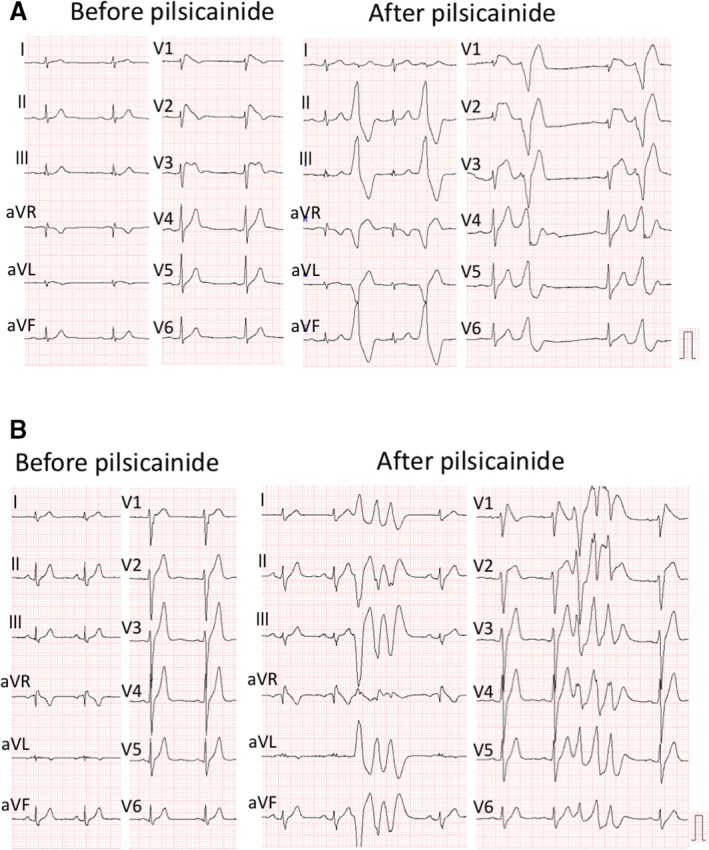
Figure 2.

Pilsicainide‐induced ventricular arrhythmia. A, These ECGs were recorded in a patient with syncope (50 years old). The left panel shows ECG at baseline. Leads V1‐2 were located at the third intercostal space. The patient had spontaneous type 1 ECG only in the leads at high intercostal spaces. The right panel shows that pilsicainide provoked frequent occurrence of premature ventricular contractions and significant ST elevation. B, These ECGs were recorded in an asymptomatic patient (27 years old). The patient had fever‐induced type 1 ECG but did not have spontaneous type 1 ECG. The left panel shows non–type 1 ECG before the pilsicainide test. Leads V1‐2 were recorded at regular lead positions. The right panel shows that pilsicainide induced nonsustained polymorphic ventricular tachycardia. The patient died suddenly at night 6 years after the test.
