Figure 6.

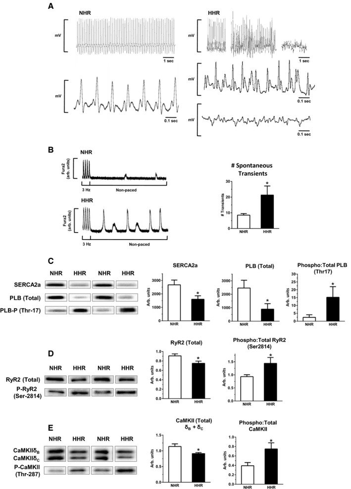
HHR in vivo and in vitro dysrhythmic substrates—cardiomyocyte Ca handling instability. A, Exemplar ECG records depicting stable sinus rhythm in normal heart rat (NHR) vs episodic hypertrophic heart rat (HHR) dysrhythmic periods. B, Spontaneous Ca2+ release events in nonpaced cardiomyocytes, quantified over 30 seconds. (*P<0.05, Student t test, n=21–23 cells/group). C through E, Immunoblot analyses of total protein expression and phosphorylation levels, left ventricular tissue homogenate, for SERCA2a, PLB (Ser‐17), RYR2 (Ser2814), CaMKII (combined δB & δC isoform, Thr287) (*P<0.05, Student t test, N=10 hearts/group). Graphs show mean ± SEM. For A, age 30 to 50 weeks. For B through E, age 20 to 30 weeks.
