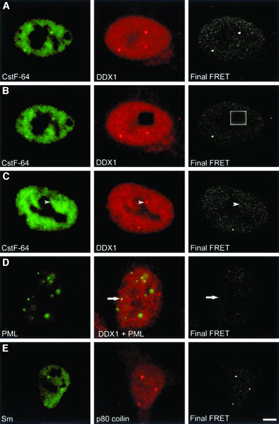
Figure 4.
DDX1 and CstF-64 undergo FRET in HeLa cells. HeLa cells were indirectly immunolabeled with anti-DDX1 antibody (2923) and either anti-CstF-64 (A–C) or anti-PML (D) antibody. The images were collected using the three-filter set method as described in MATERIALS AND METHODS. The first column represents the image obtained with the donor filters. The image in the second column was collected with the acceptor filters (except D, which represents the combined acceptor and donor signals). The third column represents the final FRET image processed to remove non-FRET components (as described in MATERIALS AND METHODS). (A) FRET was observed between CstF-64 and DDX1 in both the nucleoplasm and cleavage bodies. (B) After photodestruction of the acceptor molecule, FRET no longer occurred (boxed region). (C) Areas lacking CstF-64 foci (arrowhead) often did not demonstrate FRET. (D) Another subnuclear body, PML, demonstrated little FRET with DDX1. The arrow indicates the location of adjacent PML and DDX1 foci. (E) FRET was observed between two components of Cajal bodies, p80 coilin and Sm proteins. Bar, 5 μm.

