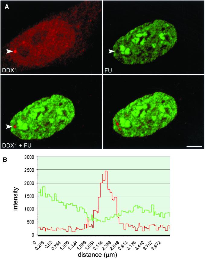
Figure 5.
DDX1 foci do not accumulate nascent RNA. (A) HeLa cells were incubated with FU for 15 min and stained with anti-DDX1 antibody (2923) and anti-bromodeoxyuridine antibody (FU). The arrowhead indicates a DDX1 focus. (B) The staining intensities of the region through a DDX1 foci (highlighted by the arrow in the bottom left panel) were profiled with the use of the Zeiss LSM 510 image software. The green line represents FU intensity, whereas the red line represents DDX1 intensity. Bar, 5 μm.

