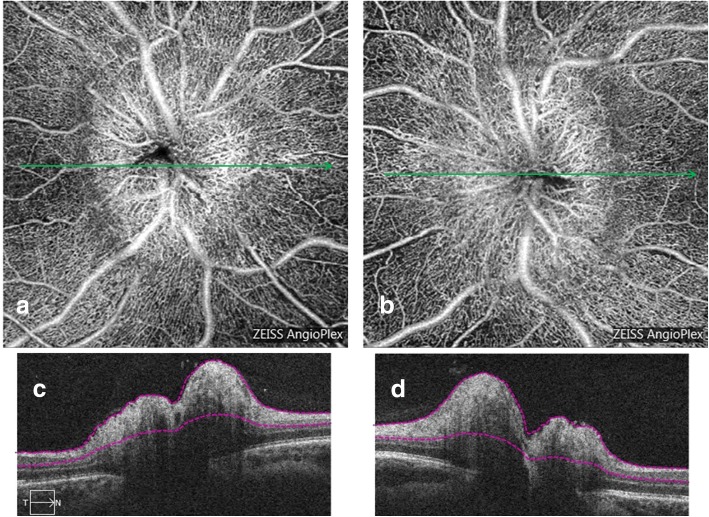
Fig. 4.
A 25 years old female presenting bilateral papilledema related to idiopathic intracranial hypertension. Superficial OCT-A of both eyes (a and b) acquired with Angioplex© demonstrating a dilation of the superficial optic disc vessels, which are tortuous and dilated. The global aspect of the optic disc looks like a tangled ball of vessels (arrowheads). OCT-B scans (c and d) corresponding to a and b

