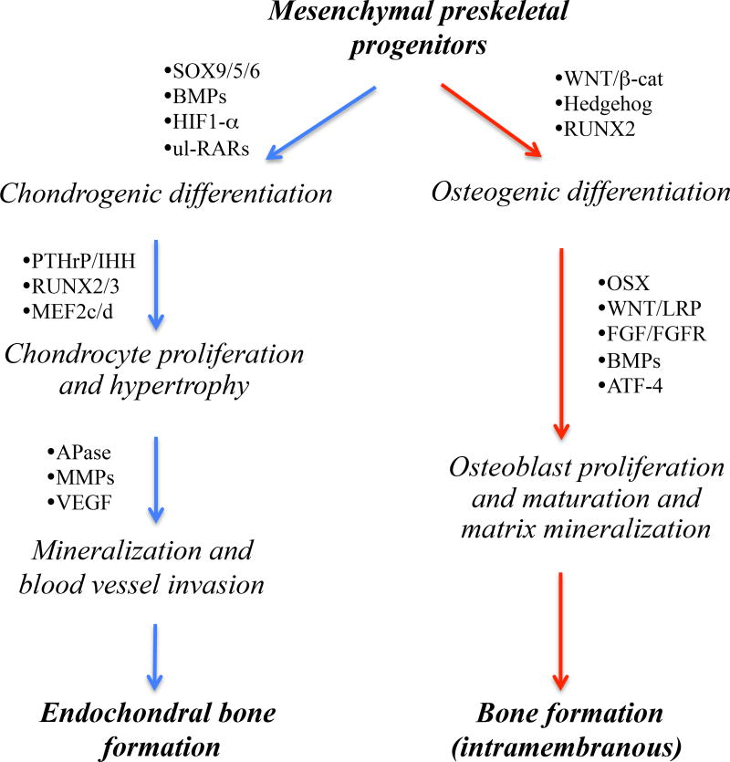
Fig. 1.
Schematic describes the major developmental steps that underlie the differentiation of preskeletal mesenchymal cells toward endochondral (right) and intramembranous (left) ossification. Multiple distinct mechanisms control fate determination and commitment of the progenitor cells to either developmental pathway, including hypoxia and retinoid and Wnt/β-catenin signaling that are particularly relevant to studies discussed in this review article. In many if not all respects, these developmental pathways are recapitulated in the formation of endochondral and intramembranous masses of HO tissue. Thus, specific steps and factors involved in each developmental pathway are being tested and exploited as possible and reasonable therapeutic targets for acquired and/or congenital forms of HO (as described in the text). Note that factors and pathways mediating the transition from mineralized hypertrophic cartilage to endochondral bone are very similar to those regulating osteogenic differentiation (listed on the left). BMP, bone morphogenetic protein; HIF, hypoxia-inducible factor; RAR, retinoic acid receptor; ul-RAR, unliganded RAR; PTHrP, parathyroid hormone-related protein; IHH, indian hedgehog; APase, alkaline phosphatase; MMP, matrix metalloprotease; VEGF, vascular endothelial growth factor; FGF, fibroblast growth factor; FGFR, fibroblast growth factor receptor; and OSX, osterix.

