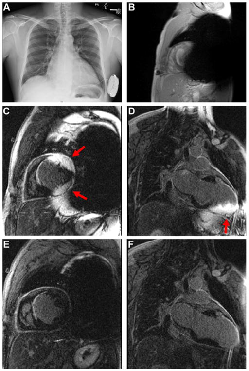
Figure 1.
(A) Chest X-ray of a 37-year old man with an MR-conditional S-ICD implanted on the left side. (B) Scout image in a short axis view the patient showing significant signal voids induced by the S-ICD. (C and D) Standard LGE images in short-axis and 2-chamber planes. (E and F) The corresponding wideband LGE images in short-axis and 2-chamber planes. Arrows point to image artifact induced by the S-ICD. MRI images were obtained with standard imaging parameters, including spatial resolution, inversion time, and gradient echo readout.

