Fig. 1.
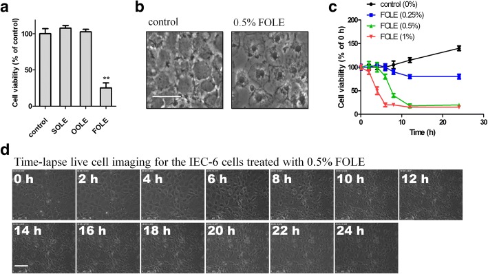
FOLE induces necrotic cell death in IEC-6 cells. a Cell viability assessed by CCK-8 assay. IEC-6 cells were treated with 0.25% SOLE, 0.25% OOLE and 0.5% FOLE for 24 h. Note that a significant reduction in the viability was observed in FOLE-treated cells. Data are presented as mean ± SD. **, p < 0.01 compared to control (n = 6–8 wells/group). b Necrotic cell death in FOLE-treated cells. Representative images are shown (Scale bar = 25 μm). c Cell viability assessed by CCK-8 assay. IEC-6 cells were treated with 0.25–1% FOLE for 0–24 h. Notably, FOLE reduced the cell viability of IEC-6 cells in a dose-dependent manner, and rapid reduction was observed at 4–8 h post FOLE treatment. d Time-lapse live cell imaging. Post-confluent IEC-6 cells were treated with 0.5% FOLE, and a 24-h time-lapse live cell imaging was performed as described in material and methods. Representative images are shown. Scale bar = 75 μm. Videos are available as supplementary data (see Additional file 1: IEC-6 cells treated with FOLE.avi). Three experiments were performed that showed similar results

