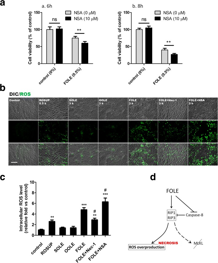
Fig. 4.
FOLE-induced necroptosis is mediated via ROS overproduction instead of MLKL. a Cell viability. IEC-6 cells were pre-treated with NSA (10 μM) for 40 min, followed by exposure to 0.5% FOLE for 6 h (a) and 8 h (b). Data are presented as mean ± SD. **, p < 0.01 (n = 6–8 wells/group). b Representative images of ROS production in FOLE-treated cells. Three hours after FOLE-treatment, the levels of intracellular ROS were determined by DCFDA as described in material and methods. Pretreatment with Nec-1 and NSA was performed as described previously. Treatment with ROSUP (30 min) served as positive control. ROS signal is presented as green fluorescence. Scale bar = 100 μm. Three experiments were performed that showed similar results. c Quantification of intracellular ROS levels. Cells were treated as described in (b). Data are presented as mean ± SD **, p < 0.01 ***, p < 0.001 compared to control; #, p < 0.05 compared to FOLE (n = 6–8 wells/group). d A schematic representation showing that ROS overproduction instead of MLKL is at least in part responsible for FOLE-induced necroptosis

