
Figure 5.
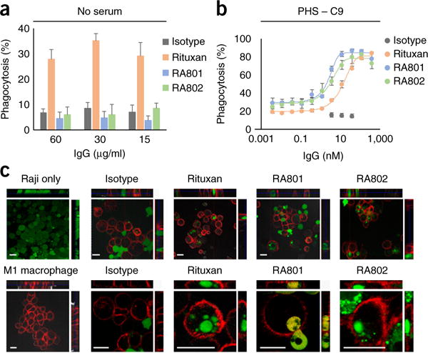
CDCP of CD20+ cells. (a,b) Phagocytosis of antibody-opsonized Raji cells (antibodies in key; concentration, horizontal axes) by monocytederived human M1-macrophages in RPMI-1640 medium without serum (a) or with serum depleted of C9 (b). (c) Fluorescence microscopy analyzing the phagocytosis of Raji cells, stained with the cell-permeant dye calcein-AM (green) by M1 macrophages, stained with anti-CD14 and anti-CD11b with allophycocyanin (red), showing Raji cells without macrophages (top left) or macrophages without Raji cells (bottom left), with opsonization by isotype-matched control antibody, Rituxan, RA801 or RA802 in the presence of serum depleted of C9 (top row, low magnification; bottom row, high magnification); top and right ‘strips’ show different views (xz and yz) of the same cells at a confocal plane. Scale bars, 20 μm. Data are from one experiment representative of three experiments (error bars (a,b), s.d. of technical triplicates).