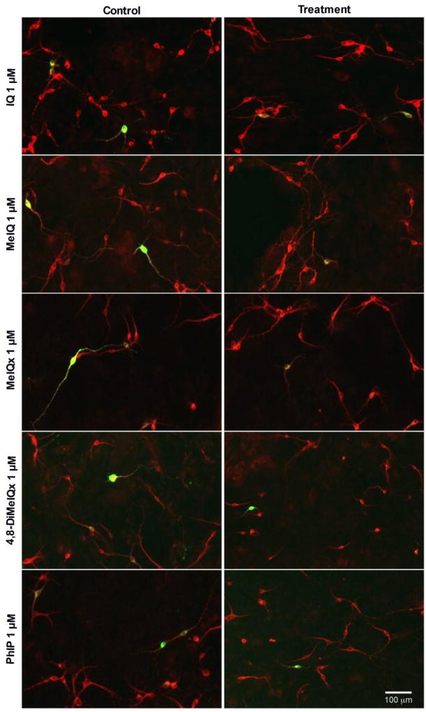
Figure 2. Representative images of primary midbrain neuronal cultures exposed to aminoimidazoaazarene HCAs (1 µM) show selective dopaminergic neurotoxicity.
Primary midbrain cultures from E17 rat embryos were treated with IQ, MeIQ, MeIQx, 4,8-DiMeIQx and PhIP for 24 h at concentrations of 100 nM – 5 µM. Cells were stained for tyrosine hydroxylase, TH (green) to identify dopamine neurons and microtubule-associated protein 2, MAP2 (red) to identify all neurons. Quantification was performed at all doses, while representative images are shown here from a 1 µM treatment, where the majority of tested HCAs exhibited selective dopamine toxicity. Images were obtained used an automated Cytation 3 cell imaging reader with a 4× objective. Scale bar represents 100 µm.

