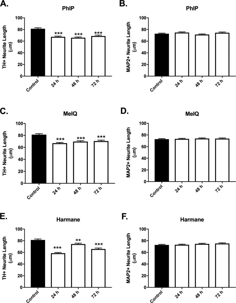
Figure 9. Temporal development of HCA-induced selective decreases in dopaminergic neurite lengths.
Decreased dopaminergic neurite lengths were detected in cultures exposed to PhIP (A), MeIQ (C), and harmane (E) at 1 µM (24 – 72 h) (n = 519 – 834 TH+ neurites/group analyzed). Non-dopaminergic neurites are not affected at the tested dose at all time-points (B,D,F) (n = 859 – 1204). Cells were stained for TH (green) to identify dopamine neurons and MAP2 (red) to identify all neurons. Data presented as the mean ± SEM; general linear model with Dunnett’s post-hoc test, **p<0.01, ***p< 0.001 compared to control. n = total number of neurites measured from 3 different experiments.

